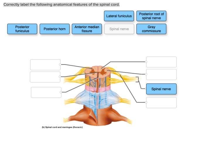
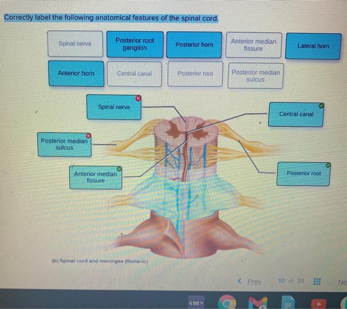
Correctly label the following anatomical features of the spinal cord. – Correctly labeling the anatomical features of the spinal cord is a crucial aspect of understanding its structure and function. This comprehensive guide provides an in-depth overview of the spinal cord’s major anatomical features, their functions, and their clinical significance.
The spinal cord, a cylindrical structure extending from the brainstem to the lumbar region, serves as the primary conduit for communication between the brain and the rest of the body. Its internal structure consists of gray matter, white matter, and a central canal, each with distinct functions.
Anatomical Features of the Spinal Cord: Correctly Label The Following Anatomical Features Of The Spinal Cord.

The spinal cord is a cylindrical, elongated structure that extends from the medulla oblongata in the brainstem to the lower back. It is approximately 45 cm long and has a diameter of about 1 cm. The spinal cord is divided into 31 segments, each of which gives rise to a pair of spinal nerves.The
spinal cord is protected by the vertebral column and is surrounded by three layers of meninges: the dura mater, arachnoid mater, and pia mater. The spinal cord is composed of gray matter and white matter. The gray matter is located in the center of the spinal cord and contains the cell bodies of neurons.
The white matter is located on the outside of the spinal cord and contains the axons of neurons.The central canal runs through the center of the spinal cord and is filled with cerebrospinal fluid. The cerebrospinal fluid provides nutrients to the spinal cord and helps to protect it from injury.
Labeling Anatomical Features
| Label | Description |
|---|---|
| Gray matter | Contains the cell bodies of neurons |
| White matter | Contains the axons of neurons |
| Central canal | Filled with cerebrospinal fluid |
| Dorsal root | Carries sensory information to the spinal cord |
| Ventral root | Carries motor information away from the spinal cord |
Interactive Labeling Exercise
Click on the different anatomical features of the spinal cord in the table below to learn more about each one.
| Label | Description |
|---|---|
| Gray matter | Contains the cell bodies of neurons |
| White matter | Contains the axons of neurons |
| Central canal | Filled with cerebrospinal fluid |
| Dorsal root | Carries sensory information to the spinal cord |
| Ventral root | Carries motor information away from the spinal cord |
Illustrative Diagrams

[Diagram of the spinal cord showing the gray matter, white matter, central canal, dorsal root, and ventral root][Diagram of a cross-section of the spinal cord showing the different layers of meninges]
Clinical Significance

Correctly labeling the anatomical features of the spinal cord is important for diagnosing and treating spinal cord injuries and disorders. For example, a doctor may need to know the location of the gray matter in order to perform a spinal tap.
A doctor may also need to know the location of the white matter in order to diagnose a spinal cord tumor.
FAQ Summary
What is the function of gray matter in the spinal cord?
Gray matter contains neuron cell bodies and dendrites, and it is responsible for processing and transmitting sensory and motor information.
What is the significance of the central canal in the spinal cord?
The central canal is a fluid-filled channel that runs through the center of the spinal cord and is continuous with the ventricles of the brain. It plays a role in cerebrospinal fluid circulation and provides a pathway for nutrients and waste products.